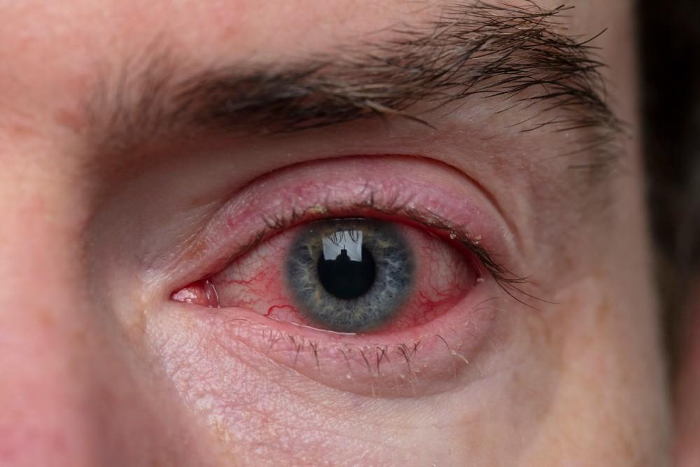
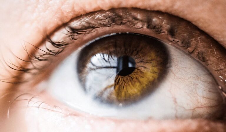
Retinal Detachment Treatments: What to Expect and When to Seek Help
Retinal detachment is a serious ocular condition that can lead to permanent vision loss if not treated promptly. Understanding the nature of this condition, its symptoms, diagnosis, and available treatments is crucial for both prevention and effective intervention. This article aims to provide comprehensive insights into retinal detachment, outlining what to expect through the entire process from symptoms to recovery.
Understanding Retinal Detachment
Retinal detachment occurs when the retina, a thin layer of tissue at the back of the eye, separates from its underlying supportive tissue. This separation deprives the retinal cells of vital nutrients and oxygen, which can ultimately result in vision loss. Understanding its nature is vital for anyone at risk or experiencing related symptoms.
What is Retinal Detachment?
Retinal detachment teatements is categorized primarily into three types: rhegmatogenous, tractional, and exudative. Rhegmatogenous detachment is the most common and results from a tear in the retina, allowing fluid to enter underneath. Tractional detachment occurs when fibrous tissue pulls the retina away from its underlying layer, while exudative detachment results from fluid accumulation beneath the retina due to various diseases.
The retina is crucial for processing visual information, and its detachment can lead to significant visual impairment. When retinal detachment occurs, immediate medical attention is essential to increase the chances of preserving sight. Symptoms may include sudden flashes of light, floaters, or a shadow over the visual field, which should never be ignored. Early intervention can often lead to successful surgical repair, restoring vision and preventing further complications.
Causes and Risk Factors of Retinal Detachment
Several factors can contribute to the risk of developing retinal detachment. Age is a significant factor, especially individuals over 50 are more susceptible due to natural degenerative changes in the eye. Other risk factors include high myopia (nearsightedness), a family history of retinal detachment, previous eye surgeries or injuries, and certain medical conditions like diabetes.
Understanding these causes and risk factors can aid in early detection and preventive measures. Regular eye examinations are especially critical for those with known risk factors. Additionally, individuals who have undergone cataract surgery or have experienced trauma to the eye should be particularly vigilant. Awareness of the symptoms and maintaining open communication with an eye care professional can help ensure timely intervention, which is crucial for preserving vision. Moreover, lifestyle choices such as managing systemic health conditions and protecting the eyes from injury can also play a significant role in reducing the risk of retinal detachment.
Symptoms of Retinal Detachment
Recognizing the symptoms of retinal detachment is crucial for timely intervention. The earlier the condition is diagnosed and treated, the better the outcomes for vision preservation.
Early Warning Signs
Early warning signs can manifest in various forms. Individuals may notice sudden flashes of light in one or both eyes, which may be followed by an increase in floaters—tiny specks or lines that float in your vision. Additionally, a shadow or curtain effect may appear in peripheral vision, indicating a possible detachment. These symptoms can often be alarming, and their sudden onset can lead to anxiety and concern about one’s eye health.
If anyone observes these symptoms, they should seek medical attention immediately, as prompt treatment is often critical for stabilizing the condition. It is also important to note that these symptoms can sometimes be mistaken for less serious eye issues, such as migraines or eye strain. However, understanding the potential severity of retinal detachment can help individuals take these signs seriously and act swiftly.
Progression of Symptoms
As retinal detachment progresses, symptoms may worsen. This can include a significant decrease in vision, where straight lines might appear distorted or wavy. One might also experience blurriness or complete loss of vision in the affected eye. These signs demand urgent medical evaluation as they may signify extensive retinal damage. In some cases, individuals may also report a sudden onset of a dark veil or shadow obscuring part of their visual field, which can be particularly distressing.
Awareness of how symptoms can progress reinforces the importance of seeking help when experiencing any unusual visual changes. Furthermore, individuals with risk factors such as high myopia, previous eye surgery, or a family history of retinal issues should be particularly vigilant. Regular eye examinations can also play a vital role in early detection, allowing for monitoring of any changes that may indicate the onset of retinal detachment, thus enabling proactive management of eye health.
Diagnosis of Retinal Detachment
Diagnosis of retinal detachment typically requires a comprehensive eye examination performed by an ophthalmologist. This process helps to establish the extent of detachment and the appropriate treatment plan.
Eye Examination Procedures
During an eye examination, an ophthalmologist may perform several tests, including visual acuity tests to assess overall vision quality. Additionally, the doctor will use dilating drops to widen the pupil, providing better visibility of the retina and facilitating detailed examination.

Slit-lamp examination and indirect ophthalmoscopy are common techniques used to visualize the retina clearly. These procedures are essential for accurate diagnosis and proper treatment planning. The slit lamp allows the doctor to examine the front structures of the eye in detail, while indirect ophthalmoscopy enables a broader view of the retina, making it easier to identify any tears or holes that may indicate detachment. The combination of these methods ensures a thorough evaluation of the eye’s health.
Imaging Techniques for Retinal Detachment
In certain cases, imaging techniques such as ultrasound might be employed, especially when the view of the retina is obscured due to cataracts or bleeding. This non-invasive procedure provides clarity about the extent and type of retinal detachment. Ultrasound can also help differentiate between various types of retinal detachments, such as rhegmatogenous, tractional, or exudative, which is crucial for determining the most effective treatment approach.
Advanced imaging technologies, such as optical coherence tomography (OCT), can offer even more intricate detail about the retina’s structure, aiding the ophthalmologist in making informed decisions regarding treatment options. OCT works by capturing high-resolution cross-sectional images of the retina, allowing the doctor to visualize the layers of the retina and assess any abnormalities. This technology not only enhances diagnostic accuracy but also plays a significant role in monitoring the effectiveness of treatment over time, providing valuable insights into the healing process.
Treatment Options for Retinal Detachment
Treatment for retinal detachment depends on its type, severity, and the overall condition of the eye. Options generally range from non-surgical to surgical interventions.
Non-Surgical Treatments
Non-surgical treatments are mainly applicable in cases where the detachment is recent and not extensive. One such approach is observation, where the ophthalmologist monitors the condition without immediate intervention. In some cases, laser photocoagulation or cryopexy may be employed to reattach the retina by creating adhesions around the tear.
These non-invasive treatments can prevent further detachment and stabilize the retina while preserving vision when performed in a timely manner. Additionally, patients are often advised to avoid strenuous activities or positions that could exacerbate the condition, such as bending over or heavy lifting. Regular follow-up appointments are crucial to assess the retina’s status and ensure that any changes are promptly addressed, as early detection of complications can significantly influence the outcome.
Surgical Procedures for Retinal Detachment
In cases of severe detachment, surgical intervention may be necessary. Various procedures are available, including scleral buckling, vitrectomy, and pneumatic retinopexy.
- Scleral Buckling: Involves placing a silicone band around the eye to relieve tension on the retina and promote reattachment.
- Vitrectomy: This surgery removes the vitreous gel that pulls on the retina, allowing the retina to flatten out and reattach.
- Pneumatic Retinopexy: Involves injecting a gas bubble into the eye, which pushes the retina back into position.
The choice of procedure is based on the individual case and the eye surgeon’s recommendation. Each surgical option comes with its own set of risks and benefits, and the decision-making process often includes a detailed discussion between the patient and the surgeon about the expected outcomes and potential complications. Post-operative care is equally important, as patients may need to maintain specific head positions to help the retina heal properly, and follow-up visits are essential to monitor the success of the surgery and detect any signs of re-detachment or other issues.
What to Expect During and After Treatment
Understanding what to expect during and after treatment can alleviate concerns and enhance the experience of patients undergoing retinal detachment procedures.
During the Procedure
Most retinal detachment surgeries are performed under local anesthesia, and patients are typically awake during the procedure. They may experience pressure sensations during surgery but no pain should be felt. The surgical team will guide patients through each step, keeping them informed to minimize anxiety.
Length of the procedure varies according to the method used, but generally ranges from one to three hours. Following the surgery, patients are monitored in a recovery area before being discharged. It’s essential for patients to have a trusted companion ready to take them home, as the effects of anesthesia can linger, making it unsafe to drive immediately after the procedure. This time also allows patients to ask any lingering questions about their recovery and care instructions.

Recovery and Post-Operative Care
Recovery from retinal detachment surgery involves specific post-operative care, including avoiding strenuous activities and head positioning as recommended by the surgeon. Follow-up appointments will be critical to monitor healing and ensure the retina remains securely attached. Patients may also be advised to use eye shields or glasses to protect their eyes during the initial recovery phase, which can last several weeks.
Vision may take time to stabilize, and patients should be aware that some fluctuations may occur during the recovery process. Adhering to post-operative instructions is vital, as it significantly impacts the overall success of the treatment. Additionally, patients might experience some discomfort, such as mild irritation or tearing, which is normal. It’s important to report any sudden changes in vision or increased pain to the healthcare provider, as these could indicate complications. Emotional support during this time can also be beneficial, as the uncertainty of recovery can be stressful. Engaging in light activities, such as reading or listening to music, can help distract from the healing process while still allowing for rest and recovery.
In summary, understanding retinal detachment, its symptoms, diagnosis, and treatment is essential for preserving vision. Individuals experiencing any warning signs should seek immediate help, as timely intervention can lead to better outcomes and successful recovery.
See Also: Signs of retinal damage: when to see a retinal specialist.


